
Contents
What is a PET scan?
Positron Emission Tomography (PET) scans utilize a radioactive material known as a tracer; they examine the body for abnormalities.
It can be used to monitor
- The function of tissues and organs can be seen with a PET scan.
- These examinations display the internal structure and blood circulation to and from the organs.
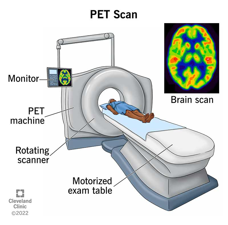
How do PET scans work?
PET detects photons—subatomic particles—emitted by a radionuclide in the organ or tissue under examination via a scanning device.
Process of PET scan
The radionuclides used in this scans are formed when the radioactive atom is combined with chemical substances that the particular organ or tissue naturally uses during its metabolic process.
For instance, because the brain uses glucose for metabolism, a radioactive atom is added to glucose to produce the radionuclide fluorodeoxyglucose (FDG) in PET scans of the brain. Thus, a common technique in PET scanning is FDG.
Depending on the scan’s aim, several materials may be utilized for this scanning. If the blood flow and perfusion of a particular organ or tissue are of interest, a radioactive form of oxygen, carbon, nitrogen, or gallium may be used as the radionuclide
During the scan
A healthcare provider injects the radionuclide into a vein using an intravenous (IV) line. The scanner then slowly passes over the body portion under examination. As the radionuclide breaks down, it releases positrons. When positrons collide with electrons, they produce gamma rays known as annihilation photons.
Then, when the annihilation photons happen to arrive at the detectors 180 degrees apart by coincidence, the scanner detects them.
Image formation
A computer analyzes these gamma rays and uses the data to produce an imaging map of the tissue or organ under study. Thus, the level of radioactivity accumulated in the tissue determines the degree of organ or tissue function and impacts the brightness of the tissue on the image.
Purpose of PET Scan
- This scans are generally useful for assessing the presence of medical conditions or different diseases in organs and/or tissues.
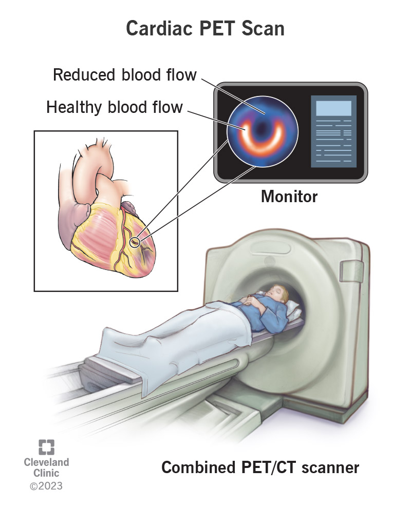
- Doctors use it to assess how well organs like the heart or brain are functioning.
- Doctors use it to diagnose and assess cancer therapies.
Doctors diagnose neurological disorders like Alzheimer’s disease and other dementias
- Parkinson’s disease
- Huntington’s disease
- Epilepsy
- Cerebrovascular accident (stroke)
Other Diagnosis
- To determine the precise surgical site before performing brain surgery.
- Examining the brain following trauma to find any bleeding, hematomas, or abnormalities in the blood or oxygen flow to the brain tissue.
- To determine if the initial cancer site has spread to other parts of the body with malignancy.
- To assess how well cancer treatments work.
- Also, to assess the effectiveness of a treatment therapy to increase blood flow to the myocardial (heart muscle) by measuring the perfusion (blood flow) to the myocardium.
- To more precisely identify lung tumors or lesions found on a chest CT scan or X-ray.
- To support the staging of lesions and monitoring of lesions following treatment in order to aid in the management and treatment of lung cancer.
Contrast Used for PET Scan
Tissues and cells utilize a material with a tracer attached to it, but cancer cells do not use the substance in the same way as healthy cells. The healthcare professional can observe how cells are utilizing the material due to the radioactive component of the tracer. Thus, they can discover cancer cells.
PET-CT will use FDG as the tracer. You will get the tracer through a catheter. Then, the catheter may be an intravenous (IV) line in your hand or arm, or it may be a central venous catheter (CVC). Cells use the tracer, which the body quickly eliminates. Urine is the primary way that it exits the body.
FDG PET Scan
Cancer cells need more glucose than healthy cells to develop and spread. As a result, cancerous regions of the body will accumulate FDG radiotracers.
How does it work?
- The patient receives an injection of FDG radiotracer into the vein.
- This scan finds FDG and creates pictures of the patient’s organs.
- Bright areas are indicative of cancer on PET imaging. It’s typical for some organs to appear bright as well.
- Doctors can infer the probability of a cancer diagnosis from the patient’s symptoms, the brightness, and the shape of the spots
FDG PET uses
- Recognize a lump or growth if they believe it to be a new cancer diagnosis or the reappearance of an earlier malignancy.
- To ascertain the cancer’s stage, search for distant cancerous regions or metastases. The stage of a malignancy may affect available treatments.
- Doctors use it to examine the progress of cancer treatment.
Conventional imaging techniques for lymphoma detection
CT Scans
Previously, CT scans have been a dependable method for lymphoma staging, offering high sensitivity and specificity prior to treatment by identifying the location and size of tumors. However, they have low specificity post-therapy, as they can’t distinguish between viable tumors and non-cancerous tissue like necrotic or scar tissue. Thus, the limitation often leads to residual masses being detected, which may not indicate persistent disease.
Gallium Scanning
Gallium-67 scintigraphy improves the specificity of CT scans by using a metabolic study to detect viable lymphoma cells, with high sensitivity and specificity in distinguishing lymphoma from benign tissue.
Despite its benefits, gallium scanning is less effective for indolent lymphomas and abdominal disease due to low spatial resolution and the time-consuming nature of the scans. Inconsistent imaging approaches and the need for advanced techniques like SPECT further limit its usage.
PET/CT Scan
PET/CT combines a multidetector helical CT scan and a full-ring detector PET scanner, allowing them to obtain the PET scan right after the CT scan. The fusion of the images precisely localizes the aberrant lesions. Also, PET/CT offers more sensitive and specific imaging. It also produces attenuation-corrected PET pictures much faster than when they combine emission and transmission PET scans. Thus, the scans are rapidly replacing PET scanners.
It is an important advancement in non-invasive lymphoma assessment. In patients with Hodgkin lymphoma (HL) and the majority of slow and severe NHL subtypes, it exhibits great sensitivity and specificity.
Problems
PET/CT uses
- Pretreatment staging,
- Restaging,
- Therapy monitoring,
- Posttherapy surveillance,
- Assessment of transformation in lymphoma patients.
In the US, the Center for Medicare and Medicaid Services (CMS) essentially reimburses PET scans for staging, restaging, and evaluating the transition from latent to serious NHL; however, using it for therapy monitoring and post-therapy surveillance is still pending approval.
Advantages of PET Scan
- PET can identify aberrant tissues, which may be more or less active than normal tissues, and provide information about the function of the tissue.
- High sensitivity and specificity.
- The capacity to identify problems early on.
- Non-invasive nature and the capability to provide functional information.
Risks of PET Scan
- PET is not as good as computed tomography (CT) or magnetic resonance imaging (MRI) for displaying the anatomical and structural details of tissues and organs.
- Patients may be instructed to stay away from close contact with infants, young children, and pregnant women for a few hours following a PET scan as a precaution. The reason for this is that the patient was mildly radioactive at the time.
- The CT portion of a PET-CT scan exposes patients to a small amount of additional radiation, but it carries very little risk of causing future issues.
Application of PET Scan
- Clinicians widely use PET scans to detect cancer, assess its spread, and stage the disease.
- It helps to evaluate how well cancer treatments, such as chemotherapy or radiation, are working by monitoring changes in metabolic activity.
- It can detect the recurrence of tumors by highlighting areas of abnormal metabolic activity that might indicate cancer’s return.
- It is used to diagnose and manage neurological conditions like Alzheimer’s disease, epilepsy, and Parkinson’s disease by visualizing brain function.
- It can determine areas of decreased blood flow, measure heart function, and determine whether cardiac tissue is still viable following a heart attack.
- Thus, it helps in guiding biopsies and surgical procedures by precisely locating abnormal tissues that need to be sampled or removed.
- Also, clinicians use it in research to examine underlying disease processes and evaluate the effectiveness of new drugs.
Sources
- https://jamanetwork.com/journals/jamaoncology/fullarticle/2807033
- https://www.mskcc.org/cancer-care/patient-education/pet-ct-fdg#:~:text=You%20will%20get%20oral%20contrast,contrast%20(contrast%20with%20iodine)
- https://ashpublications.org/blood/article/110/10/3507/23359/The-role-of-FDG-PET-scans-in-patients-with
- https://www.msdmanuals.com/en-in/home/special-subjects/common-imaging-tests/positron-emission-tomography-pet
- https://www.nhs.uk/conditions/pet-scan/#:~:text=Possible%20risks%20of%20a%20PET,standard%20PET%20scan%20is%20safe
- https://my.clevelandclinic.org/health/diagnostics/10123-pet-scan
- https://my.clevelandclinic.org/health/diagnostics/17376-cardiac-positron-emission-tomography-pet
Written by Snegkha S