
Contents
What are lymphoproliferative disorders?
Lymphoproliferative disorders (LPD) consist of a variety of diseases. Basically, lymphocytes are produced in an uncontrolled manner in these disease. Generally, there are two types of lymphocytes in blood: B lymphocytes and T lymphocytes. LPD leads to monoclonal lymphocytosis, lymphadenopathy, and bone marrow infiltration. Consequently, the lymphocytes proliferate in an uncontrolled manner, leading to immunodeficiency, a defective immune system, and lymphocyte imbalance.
Types of lymphoproliferative disorders
Chronic lymphocytic leukemia (CLL)
Chronic lymphocytic leukemia is one of the common types of leukemia. It generally affects people over the age of 65, but it can also affect people in their 30’s. Consequently, an abnormal increase in defective lymphocytes is characteristic of this disease. Usually, these defective lymphocytes are called leukemia cells.
Based on the cells they affect, CLL can be of two types. If B cell proliferation exceeds the normal level, it results in B cell Chronic Lymphocytic Leukemia . Similarly, the condition is known as T cell CLL, if the T cells are affected. Those with T cell CLL experience earlier of symptoms than those with B cell CLL.
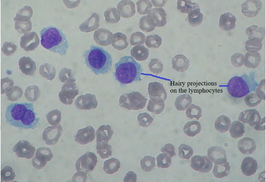
Hairy cell leukemia
Hairy cell leukemia is chronic, rare B cell leukemia. Under a microscope, hair-like projections appear to cover the lymphocytes in this disease. Usually the spleen, bone marrow, and blood all harbor cancer cells. As the disease progresses slowly, it can be kept under control if treated properly.
Waldenstrom’s macroglobulinemia
Lymphoplasmacytic lymphoma is also known as Waldenstrom’s macroglobulinemia. It is a rare form of B cell lymphoma. It usually progresses slowly. In this disease, the number of monoclonal IgM antibodies and M proteins increases considerably in the blood. As a result of this increase in the M proteins, the thickness of the blood increases. As a result, the flow of blood in the blood vessel is reduced. This lymphoma is one form of non-Hodgkin’s lymphoma.
Mantle cell lymphoma (MCL)
Mantle cell lymphoma is one form of non-Hodgkin’s lymphoma. It is an aggressive B cell lymphoma. The mantle area of the lymph node is the origin place of mantle cell lymphoma. Lymph nodes are present throughout the body, and they play a major role in fighting against infections. This lymphoma is present in the spleen, gastrointestinal tract, bone marrow, and bloodstream.
Follicular lymphoma
One of the most prevalent forms of lymphoma is follicular lymphoma. It is a type of non-Hodgkin’s lymphoma. It is the lymphoma of the B lymphocytes. The abnormal B cells form clusters in the lymph nodes, called follicles. As a result this lymphoma is called follicular lymphoma. This lymphoma is classified into three stages, depending on the presence of large lymphomas and Grades N, 0, and 3. Two parts, 3A, and 3B, can be subdivided from the 3rd grade. Grades 1, 2 ,and 3A are assigned for slow lymphomas, while fast lymphomas are categorized under grade 3B.
Diffuse large B cell lymphoma (DLBCL)
Diffuse large B cell lymphoma is one of the most common forms of non-Hodgkin’s lymphoma. This rapidly growing lymphoma can be easily cured despite its presence in lymph nodes and various organs of the body, including the spleen, gastrointestinal tract, brain, lungs, liver, bone marrow, and others.

The lymphoma stages are based on the number of affected lymph nodes and organ involvement, with a total of four stages. The stages are
- Stage 1: A single lymph node or a group of adjacent lymph nodes is involved in this stage.
- Stage 2: Two or more lymph nodes are involved on the same side of the diaphragm.
- Stage 3: The lymph nodes from both sides of the body are involved, as are the lymph nodes above the diaphragm and the spleen.
- Stage 4: Lymphoma is widespread, involving lymph nodes, spleen, and other organs like the liver and spleen.
Hodgkin’s lymphoma
This is a relatively rare form of lymphoid neoplasia that has a high chance of recovery. Initially, this disease was known as Hodgkin’s disease. One of the main features of this disease is the presence of mononuclear Hodgkin cells and multinucleated Reed-Sternberg cells. These cells can be observed among inflammatory non-cancerous cells. This lymphoma initially appears in the cervical lymph node. The T cells surround the lymphoma cells. Two categories exist for Hodgkin lymphoma.
- Classic Hodgkin’s lymphoma
This category is divided into further categories.
- Nodular sclerosis Hodgkin’s lymphoma: It starts in the lymph nodes of the neck and chest.
- Lymphocyte rich Hodgkin’s lymphoma: People with HIV infections often develop this lymphoma. It affects the lymph nodes present in the upper body.
- Mixed cellularity Hodgkin’s lymphoma: It usually occurs in few lymph nodes of the upper body.
- lymphocytes – depleted Hodgkin’s lymphoma: It usually occurs in older people with HIV. very aggressive form of lymphoma. This lymphoma primarily affects the lymph nodes in the abdomen, spleen, liver, and bone marrow. It is often diagnosed when it has reached an advanced stage.
- Nodular lymphocyte predominant Hodgkin’s lymphoma
This lymphoma is not common. The popcorn-like appearance of lymphoma cells is what earns them the name popcorn cells. The lymph nodes of the neck and arm are the places where the lymphoma starts.
Non-Hodgkin’s lymphoma
Malignant growth in the lymph nodes is a symptom of non-Hodgkin’s lymphoma. Mantle cell lymphoma and follicular lymphoma are examples of non-Hodgkin’s lymphomas. Diffuse large B cell lymphoma, primary CNS lymphoma, marginal zone lymphoma, and Brukitt’s lymphoma. Based on the rate of growth and spread, non-Hodgkin’s lymphoma can be of two types
- Indolent: Spreads slowly and the symptoms are very few.
- Aggressive: spreads quickly, and the symptoms are severe.
Causes and risk factors of lymphoproliferative disorders
Several mutations, which can be acquired or caused by treatment or illness, consequently cause LPDs. In general, the mutation of X chromosome causes X-linked LPD. The mutation is on the long arm of the X chromosome at position 25, where the mutation results in the deletion of the SH2D1A gene. As a result, the natural killer cells and T lymphocytes are affected. Subsequently, this leads to LPD of these cells. This condition is expressed as X-LPD-1.
The mutation of the Fas gene leads to the condition called autoimmune lymphoproliferative syndrome (ALPS). This gene mutation occurs in the long arm chromosome 10.
A translocation in chromosomes 11 and 14 results in the overexpression of the gene CCND1, which is responsible for cyclin D1 production. This translocation results in mantle cell lymphoma.
Symptoms of lymphoproliferative disorders
Some of the symptoms of lymphoproliferative disorders are
- Fatigue
- Swollen lymph nodes
- Fever
- Infection
- Bleeding
- Easy bruising
- Petechiae
- Weight loss
- Pain in ribs
- Feeling early satiation
- Night sweating
- Shortness of breath
Diagnosis of lymphoproliferative disorders
An array of tests are available for diagnosing lymphoproliferative disorders. Some of the tests are
Physical examination and medical history
Medical history gives an insight into the patient’s health conditions, the symptoms they are suffering, and also the infection they have encountered. During a physical examination, the doctor can identify any changes in the body caused by the disease condition, such as an enlarged spleen, bruising, and pale skin.
Complete blood count with differential
This test counts the number of blood cells present in the blood sample. It also takes into account the morphology of the cells. The researcher identifies and reports any variation in cell count and morphology.
FISH
In the laboratory, we tag DNA with a fluorescent dye during the Fluorescent in situ hybridization procedure. This DNA is complementary to the DNA recovered from the patient. The fluorescent-tagged DNA binds with the DNA from the patient sample through complementary base pairing. The fluorescent dye will glow, showing the presence of the specific DNA. Thus, using this procedure we can identify mutations in genes.
Gene mutation test
There are many tests available to detect the presence of mutation. Generally, almost all lymphoproliferative diseases are caused by gene mutations. Thus, identifying the type and the location of the mutation helps in identifying the disease condition.
Biopsy
The medical team collects samples from bone marrow, spleen, liver, and lymph nodes for biopsy and detects the presence of mutation or gene abnormality.
Imaging
Imaging techniques such as the CT (computed tomography) scan and the PET (positron emission tomography) scan are used to identify tumors.
Treatment of lymphoproliferative disorders
The treatment for lymphoproliferative disease can be any one of the following or a combination of them depending on the severity of the disease condition.
Chemotherapy
Chemotherapy is the use of medicines or drugs to kill cancer cells. One of the most commonly used treatments for cancer kills fast-growing cancer cells. In this process, some healthy cells are also destroyed; consequently, this occurs as a result of the medicines or drugs used.
Radiation
It is the use of ionising radiation to kill the cancer cells.
At the site of tumor growth, we use beams of intense ionising radiation to kill cancer cells.
Immunotherapy
This method uses the body’s immune system to fight against cancer. Subsequently, these cells are activated or modified to fight against cancer. There are many different types of immunotherapies available for the treatment of cancer.
Stem cell transplant
In this therapy, we collect and freeze the suitable donor’s stem cells. Initially, the patient undergoes intense chemotherapy to kill cancer cells. After the chemotherapy, the patient is given defrosted stem cells; consequently, this treatment aims to facilitate recovery.
Target therapy
The therapy identifies and kills cells with the cancer-causing mutation using monoclonal antibodies or small molecule drugs. These substances identify specific proteins on cancer cells and attach to them, releasing the drug and causing the cancer cells to die.
Reference
- https://www.ncbi.nlm.nih.gov/books/NBK537162/#:~:text=Lymphoproliferative%20disease%20(LPD)%20is%20a,that%20can%20often%20be%20fatal.
- https://www.cancerresearchuk.org/about-cancer/hairy-cell-leukaemia/about
- https://www.ncbi.nlm.nih.gov/books/NBK537162/
- https://www.ncbi.nlm.nih.gov/books/NBK499845/
- https://www.webmd.com/cancer/lymphoma/waldenstrom-macroglobulinemia-overview
- https://www.mayoclinic.org/diseases-conditions/waldenstrom-macroglobulinemia/symptoms-causes/syc-20359967#:~:text=Waldenstrom%20macroglobulinemia%20
- https://lymphoma.org/wp-content/uploads/2023/10/LRF_Understanding_Lymphoma_Mantle_Cell_Lymphoma_Fact_Sheet.pdf
- https://www.ncbi.nlm.nih.gov/books/NBK536985/
- https://lymphoma-action.org.uk/sites/default/files/media/documents/2023-09/Follicular%20lymphoma%20information%20PDF.pdf
- https://lymphoma.org/wp-content/uploads/2023/10/LRF_Understanding_Lymphoma_Diffuse_Large_B_Cell_Lymphoma_Fact_Sheet.pdf
- https://www.lymphoma.org/understanding-lymphoma/aboutlymphoma/nhl/dlbcl/
- https://www.cancer.org/cancer/types/hodgkin-lymphoma/about/what-is-hodgkin-disease.html
- https://www.ncbi.nlm.nih.gov/books/NBK499969/
- https://www.cancer.gov/types/lymphoma/patient/adult-nhl-treatment-pdq#:~:text=Non%2DHodgkin%20lymphoma%20is%20a,risk%20of%20non%2DHodgkin%20lymphoma.
- https://www.ncbi.nlm.nih.gov/books/NBK559328/
Written by Krishnambal.S
Pingback: Interpretation of results - CBC and Differential counts - HK Technical PGIMS