
Contents
Overview
A complete blood count (CBC ) test measures all the blood components: the red blood cells (RBC), white blood cells (WBC), and platelets. This test also takes into consideration the appearance of the cells. Consequently, this not only indicates the person’s health but also reflects the status of his immune system.
Sample collection
For the complete blood count test, they collect blood. Specifically, they collect blood from the veins. Additionally, fasting is not required for the test. Subsequently, the laboratory professional will insert a needle in the vein and collect the blood in a tube. Usually, they coat the tubes with ethylene diamine tetraacetic acid (EDTA) prior to collecting blood in them. This, in turn, prevents the blood from clotting. Afterward, they process the blood samples, and then they make slides, stain them, and examine them under a microscope.
Components of the CBC test
Blood consists of two components: the liquid component and the cellular component. The liquid component of blood is serum or plasma. The cellular components are the red blood cells (RBC), white blood cells (WBC), and platelets.
Red blood cell (RBC)

The red blood cells (RBC) constitute the majority of the cellular components of blood. Specifically the red colour of RBC is due to the presence of an iron-containing protein called haemoglobin. This haemoglobin is responsible for carrying oxygen to all parts of the body. The bone marrow is the site of RBC production. An RBC can live for 120 days on average.
- Red blood cell count: this test determines how much RBC is present in the blood.
- Haemoglobin (Hgb): measures the amount of haemoglobin or iron-containing protein in the blood.
- Hematocrit (Hct): it gives the comparison of RBCs to other cells present in blood. The hematocrit value is calculated using the formula given below:
HCT=RBC✕MCV10
- Red blood cell indices
- Red cell distribution width (RWD): it indicates the variation in the size of red blood cells.
- Mean corpuscular volume (MCV): The mean corpuscular volume or MCV measures the average size of the red blood cells. MCV is computed using the formula given below.:
MCV=HctRBC
- Mean corpuscular haemoglobin (MCH): this test measures the average amount of haemoglobin in blood. The following formula is used to calculate the MCH value:
MCH=HgbRBC
- Mean corpuscular haemoglobin concentration (MCHC): this test measures the concentration of haemoglobin in the packed RBCs. The formula given below is used to calculate MCHC:
MCHC=HgbHct
White blood cells
White blood cells (WBC) are a part of our immune system. In general, while circulating in the blood, the WBC effectively defends the body by destroying any pathogens it encounters in the blood. There are three types of WBC, based on the structure of the nucleus, the granules present in them, and their staining characteristics. They are granulocytes, lymphocytes, and basophils.
- White blood cell count: This test estimates the count of white blood cells in the patients blood.
- White blood cell differential: this estimates the number of different types of WBC,
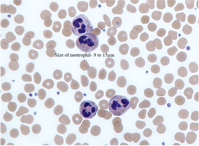
Neutrophils
Neutrophils constitute the majority of the WBC.They are granulocytes. Usually neutrophils stain a neutral colour when stained with the hematoxylin and eosin stain. They stain pink in colour. Under the microscope, the characteristic lobbed structure of the nucleus in neutrophils is clearly visible. These cells have lobed nucleus. Generally 3 – 5 lobes are present. Thus, neutrophils are also called polymorphic neutrophils.
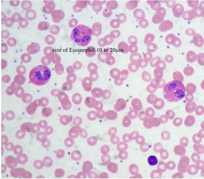
Eosinophils
It retains the colour of the acidic dye eosin when stained with hematoxylin and eosin stain. Specifically, they stain from orangish red to bright yellow in colour. Therefore, this gives rise to the name eosinophils. Additionally, eosinophils are also granulocytes.. The nucleus in eosinophils has two lobes. The cytoplasm of eosinophils has large granules. These granules have many enzymes and proteins in them.
Basophils
Basophils are the WBC responsible for allergic reactions. Specifically, they stain with the basic dye hematoxylin when stained with hematoxylin and eosin stain; consequently, they stain blue in colour. The nucleus of basophils is bi lobes and is ‘s’ shaped. The cytoplasm has many granules filled with histamine and other enzymes and proteins.

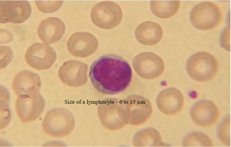
Lymphocytes
They are the second most common form of WBC. The T cells and B cells, which are the major players of the immune system, are also lymphocytes. The nucleus is spherical in shape and fills the whole cell. The cytoplasm does not have any granules. They stain blue to dark purple.
Monocytes
The number of monocytes present in the blood is very low. The nucleus of a monocyte is in the shape of a kidney bean or oval. They have an irregular appearance. The basophils stain blue in colour.

Atypical lymphocytes:
Atypical lymphocytes differ from normal lymphocytes in their shape and size. The activated lymphocytes are atypical lymphocytes. Generally these atypical lymphocytes are present during an infection.

Platelets
Platelets are the component of the WBC that helps in the clotting of the blood and prevents bleeding. Furthermore, they do not have a nucleus. However, they do contain mitochondria. The platelets are not typical cells. They are fragments from the megakaryocyte cytoplasm. These megakaryocytes are present in the bone marrow and lungs. Under the microscope, the platelets are blue or purple in colour.

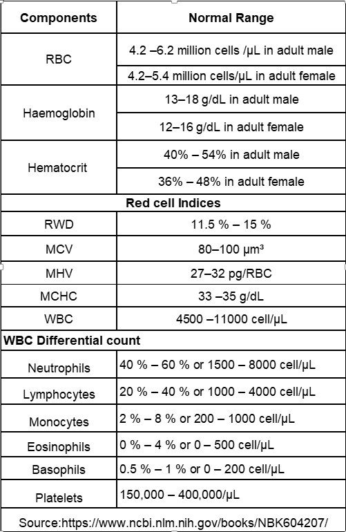
Normal range
For interpretation of results click here
Reference
- https://www.ncbi.nlm.nih.gov/books/NBK604207/
- https://medlineplus.gov/lab-tests/complete-blood-count-cbc/#:~:text=A%20complete%20blood%20count%2C%20or,fight%20infections%20and%20other%20diseases.
- https://www.webmd.com/a-to-z-guides/complete-blood-count
- https://www.webmd.com/a-to-z-guides/complete-blood-count
Written by Krishnambal.S
Pingback: PCOS and PCOD: Causes, Symptoms, Diagnosis and Differences. - HK Technical PGIMS
Pingback: Myeloproliferative Disorders - HK Technical PGIMS
Pingback: Lymphoproliferative disorders - HK Technical PGIMS
Pingback: Types of Regular Health Checkups for Optimal Health - HK Technical PGIMS