
Contents
Nuclear Medicine imaging
A radioactive tracer is injected into the body during a nuclear medicine imaging procedure. After administering the radioactive tracer material, images are created by detecting radiation from various bodily areas. Both film and a computer are used to record the images.
The following conditions are identified via nuclear medicine imaging:
- Blood disorders.
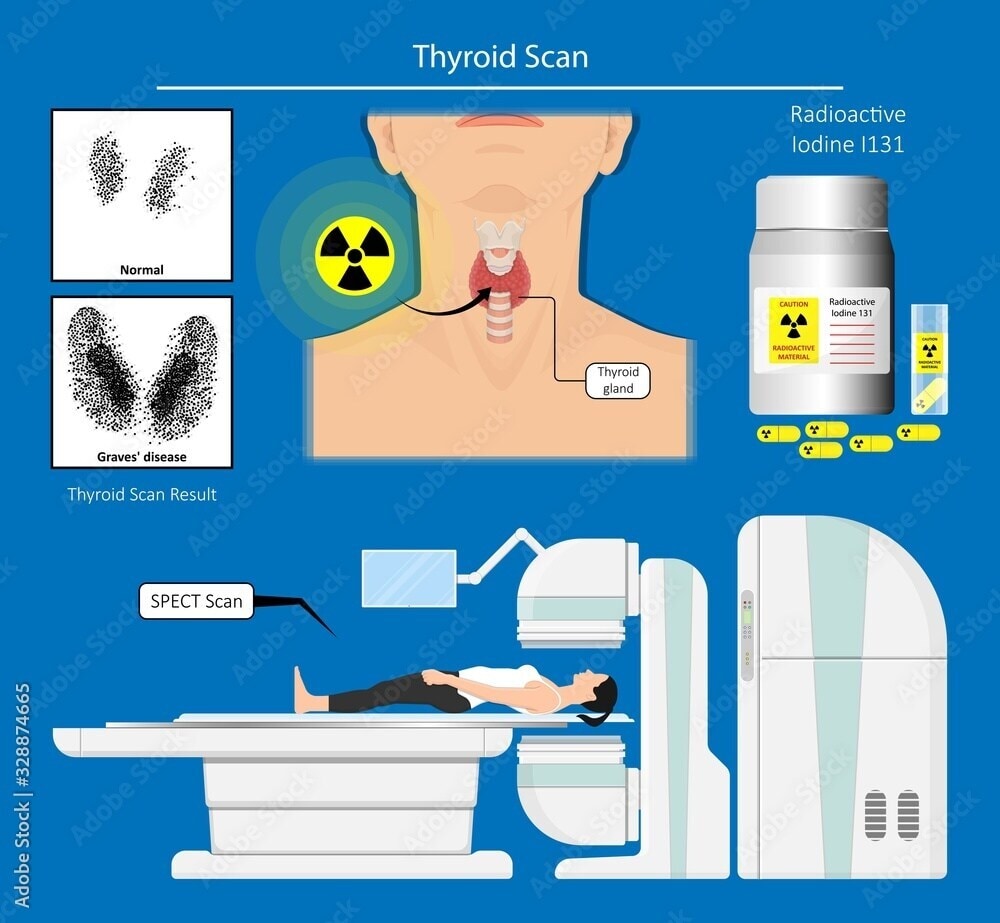
- Thyroid disease, including hypothyroidism.
- Heart disease.
- Gallbladder disease.
- Lung problems.
- Bone problems, including infections or breaks.
- Kidney disease, including infections, scars, or blockages.
- Cancer.
Imaging in nuclear medicine can also treat diseases or assess how well a treatment is functioning effectively. Radioimmunotherapy combines immunotherapy and radiation to carefully deliver radiation to a specific location.
Typically, the scans themselves take between thirty and sixty minutes, excluding the time needed for the tracer to be absorbed. Absorbing the tracer may take two or three hours in certain conditions, such as bone scans. Imaging over several days is part of a few nuclear medicine exams.
Imaging Techniques
PET (Positron Emission Tomography) in Nuclear Medicine
PET scans involve injecting a small amount of radioactive tracer, usually a form of glucose, into the body. Cancer cells, which have higher metabolic rates, absorb more of the tracer, making them visible on the scan. PET is particularly useful in oncology for detecting cancer, determining the stage of the disease, monitoring treatment response, and detecting recurrence.
PET scans assess heart function in cardiology and evaluate brain disorders like Alzheimer’s disease and epilepsy in neurology, extending beyond oncology.
SPECT (Single Photon Emission Computed Tomography)
SPECT detects gamma rays emitted by the tracer using a gamma camera, providing 3D images of the body. It is widely used in cardiology to assess coronary artery disease, evaluate heart function, and identify areas of reduced blood flow to the heart muscle.
In neurology, SPECT can help diagnose and monitor conditions like stroke, epilepsy, and brain tumors. SPECT also detects bone metastases in oncology and evaluates certain types of cancer.
Gamma Camera in Nuclear Medicine
The gamma camera is a critical component in many nuclear medicine procedures, including SPECT imaging. It detects gamma rays emitted by radioactive tracers introduced into the body.
Gamma cameras alone typically produce 2D images. The gamma camera’s crystal scintillates, or emits light, when struck by gamma rays. This light converts into electrical signals, which are then processed to create an image.
Gamma cameras perform a variety of diagnostic tests, including bone, thyroid, and renal scans, providing images to diagnose fractures, infections, tumors, and abnormal organ function. In cardiology, gamma cameras assess blood flow to the heart through myocardial perfusion imaging.
DTPA scan for kidney
A renal DTPA scan is a nuclear medicine examination in which the relative functions of the left and right kidneys are measured using a small amount of radioactive material (radioisotope). Thus, it makes it possible to locate any obstructions.
Procedure
A radioactive tracer will be injected into the patient. The patient won’t experience any change in feeling after this injection. The scan starts immediately, and the patient lies under the camera while it takes pictures for thirty minutes.
It’s possible to administer a second Lasix injection to encourage the kidney to empty. After the scan, the patient might need to use the restroom and then return for an additional five minutes of imaging.
If the word “captopril” appears in the patient referral, the patient will need to take a blood pressure medicine one hour prior to the scan, and their blood pressure will be continuously checked for the entire hour. The patient will receive a small injection of a radioactive tracer, which won’t cause any noticeable changes, followed by 20 minutes of imaging.
What is a renal DMSA scan?
There are two phases to the scan.
For the first phase, the patient will get a small injection of a radioactive tracer. They won’t experience any change in feeling after this injection. Three hours after the injection, the patient will receive a time slot from the technologist to return to the practice. The patients are allowed to engage in all regular activities during this break, such as eating, drinking, and operating a vehicle.
Patients will spend thirty minutes under the scanning camera, where it will take photographs during the second part of their visit. During this time, it is crucial to remain as motionless as possible because movement will cause the images to become blurry and lower the scan quality.
Amyloid scan
A radioisotope like fluorine F18 labels a ligand that targets Aβ plaque aggregates. As the radiopharmaceutical decays, it releases positrons. PET measures the rate of decay of these tracers in human tissue using a positron camera. NeuraceqTM (florbetaben F18), VizamylTM (flutemetamol F18), and AmyvidTM (florbetapir F18) are a few examples of these radiopharmaceuticals.
A potentially major development in the evaluation of persons with cognitive impairment is amyloid PET imaging. The scan reveals plaques in the brain, believed to cause nerve cell damage and death. Prior to the invention of amyloid PET, only brain autopsies could reveal these plaques.

Nuclear therapy for cancer treatment
Nuclear medicine therapy uses radioactive medications that bind to and kill cancer cells. The medication identifies cancerous cells. So, after intravenous injection, it circulates throughout the body, attaches itself to the tumor cells, directly exposes them to radiation, and ultimately kills them.
As part of a therapeutic approach to treat, cure, or manage the disease, nuclear medicine therapy delivers radiation to tumorous lesions by use of radiopharmaceuticals that target certain tumors, such as thyroid, lymphomas, or bone metastases. Thus, it can target specific areas or cover the entire body.
Strongly binding radiopharmaceuticals, usually referred to as vehicles with a high tumor affinity, are appropriate for therapeutic use. They can deliver precise radiation dosages straight to the metastases of cancer, protecting healthy tissue in the process. So, its affinity, or binding strength, to the target structures of the tumor, like antigens or receptors, decides which molecule delivers the radiation to the tumor. Tumors shrink as a result of the ionizing radiation that radionuclides release, which destroys cancer cells by destroying their DNA.
Radionuclides that produce ionizing radiation and have short tissue penetration, such as beta or alpha emitters, are the most suitable for tumor therapy since they release their energy close to their targets.
Various types of nuclear therapy
Thyroid Cancer
- Radioactive Iodine Therapy (I-131): This is one of the most common and successful uses of nuclear medicine. Thyroid cells, including cancerous ones, absorb radioactive iodine, which destroys them while minimizing impact on other tissues.
Prostate Cancer
- Radium-223 Therapy: This treatment targets bone metastases, which are common in advanced prostate cancer. Thus, Radium-223 mimics calcium and selectively targets bone metastases, delivering radiation directly to cancerous bone tissue.
Liver Cancer
- Radioembolization (Selective Internal Radiation Therapy, or SIRT): This therapy involves injecting radioactive microspheres into the liver’s blood supply, targeting liver tumors with high doses of radiation while sparing healthy liver tissue.
Lymphoma
- Radioimmunotherapy: This treatment combines radiation with antibodies that specifically target lymphoma cells. Thus, the antibody binds to the cancer cells, delivering a lethal dose of radiation directly to the tumor through the attached radioactive substance.

Bone Metastasis
Strontium-89 and Samarium-153: Radioactive substances treat bone pain caused by metastatic cancers, commonly spreading to the spine, pelvis, and thigh from breast or prostate cancer.
Benefits of Nuclear Medicine
- While other types of imaging analyze anatomy (the way the organs look), nuclear medicine imaging assesses how the organs function.
- So, by evaluating an organ’s function has the benefit of assisting medical professionals in diagnosing and treating the part of the body under examination.
- Thus, it can provide highly targeted treatment, which minimizes damage to surrounding healthy tissues.
Disadvantages of Nuclear Medicine
- One of the primary concerns is the potential for radiation exposure, which, although generally low, can still pose risks, particularly with repeated exposure.
- This radiation can lead to side effects such as fatigue, nausea, or even long-term risks like the development of secondary cancers.
- Moreover, the use of radioactive materials requires specialized facilities and trained personnel, which can limit the availability and accessibility of nuclear medicine treatments.
- The cost of nuclear medicine procedures can also be high, making them less accessible for some patients.
- Additionally, the effectiveness of nuclear medicine varies depending on the type and stage of the disease, and it may not be suitable for all patients.
Sources
- https://my.clevelandclinic.org/health/diagnostics/4902-nuclear-medicine-imaging
- https://snig.com.au/renal-scan-dtpa-dmsa
- https://radiology.ucsf.edu/patient-care/services/specialty-imaging/alzheimer
- https://www.cms.gov/medicare/coverage/evidence/amyloid-pet
- https://www.mayoclinic.org/departments-centers/nuclear-medicine-therapy/sections/about-nuclear-medicine-therapy/gnc-20489020
- https://www.iaea.org/topics/radionuclide-therapy#:~:text=Nuclear%20medicine%20therapy%20uses%20radiopharmaceuticals,or%20throughout%20the%20entire%20body
- https://www.cancer.org/cancer/types/thyroid-cancer/treating/radioactive-iodine.html
- https://www.cancerresearchuk.org/about-cancer/prostate-cancer/metastatic-cancer/treatment/radiotherapy/radium-223
- https://www.cancer.gov/publications/dictionaries/cancer-terms/def/radioembolization
- https://www.sciencedirect.com/science/article/abs/pii/S000129980900107X
- https://www.mayoclinic.org/diseases-conditions/bone-metastasis/symptoms-causes/syc-20370191
- https://www.sciencedirect.com/science/article/pii/S1507136710600594
Written by Snegkha S